Medical and Healthcare Machinery, MRI Machines
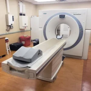
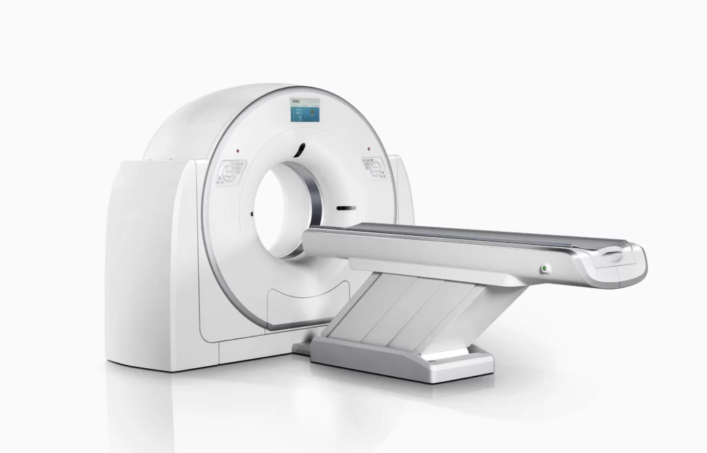
32 Slice Medical CT Scan System Machine
32-slice energy spectrum imaging technology CT enables hospitals at all levels to enjoy the advanced functions of super high-end CT. Spectral imaging platform has rich clinical value in stone composition analysis, gout examination and tumor examination.
This Machine gives a possibility to perform computed tomography without any problems and on high quality level. This device is used to conduct exams of internal organs and their functioning. With its help, a physician has a possibility to assess the condition of the human body as a whole.
Features:
- It is easy to use;
- Convenience;
- Multi functionality;
- Obtained images are of high definition;
- High-definition 3D images of the area under study;
- The procedure is pain-free;
- The data is processed fast;
- The image can be stored in the computer memory;
- The diagnostics does not take a lot of time;
- Acceptable radiation dose.
Technical Specifications:
| No. | Technical Features | Descriptions |
| 1 | Gantry | |
| 1.01 | Gantry type | Low voltage slip-ring |
| 1.02 | Gantry driven type | Strap-driven |
| 1.03 | Patient opening | 70cm |
| 1.04 | Gantry tilt mode | Digital gantry tilt |
| 1.05 | Digital tilt capability | ±50° |
| 1.06 | Detector type | OptiWave rare-earth ceramic detector |
| 1.07 | Numbers of detector rows | 16 |
| 1.08 | Width of Z-axle detector | 20mm |
| 1.09 | Detector columns of channels per row | 848 |
| 1.10 | Numbers of detector columns | 13568 |
| 1.11 | Data-transfer type | RF, optical fiber communication |
| 1.12 | Distance of focus-ISO-center | 53cm |
| 1.13 | Distance of focus-detector | 94cm |
| 1.14 | 3D laser orientation | Provided |
| 1.15 | 13″ integrated display panel | Provided |
| 1.16 | Adose automatic exposure control (mAModulation) | Provided |
| 1.17 | Auto-voice manager | Breath Graphical DisplayHold Message (Record/Playback)
Breath Message (Record/Playback) |
| 1.18 | AccuSaving energy conservation management | Provided |
| 2 | HVPS and X-ray tube | |
| 2.01 | Maximum continuous output of HVgenerator | 42kW |
| 2.02 | Tube kV selections | 70kV, 80kV, 100 kV, 120 kV, 140 kV |
| 2.03 | Tube mA range | 10~350mA |
| 2.04 | Tube anode heat capacity | 3.5MHU |
| 2.05 | Max. anode cooling rate | 735kHU/min |
| 2.06 | Type of cooling | Oil cooling + Air cooling |
| 2.07 | Tube focus | Large: 1.2mm×1.4mmSmall: 0.7mm×0.8mm |
| 2.08 | Collimator width selection | 4-level election |
| 2.09 | Focus spot tracking technology | Provided |
| 3 | Patient table | |
| 3.01 | Maximum horizontal-movable range | 1850mm |
| 3.02 | Table horizontal-scannablerange | 1800mm |
| 3.03 | Table horizontal-position repeatability | ±0.25mm |
| 3.04 | Minimum height above floor | 430mm |
| 3.05 | Maximum vertical-movable range | 500mm |
| 3.06 | Maximum speed of vertical movement | 35mm |
| 3.07 | Maximum speed of horizontal movement | 150mm/s |
| 3.08 | Maximum patient weight | 205kg |
| 3.09 | Foot pedal of patient table control | Provided |
| 4 | Computer | |
| 4.01 | CPU | 3.5GHz |
| 4.02 | Memory | 32GB |
| 4.03 | Storage of hard-disk | 1TB×2 |
| 4.04 | Monitor | 24’’ LCD Monitor |
| 4.05 | Resolution of monitor | 1920×1200 |
| 4.06 | Image-data external storage type | CD/DVD/USB |
| 4.07 | Time of image reconstruction (512×512) | 33.3ms/image |
| 4.08 | Speed of image reconstruction (512×12) | 30fps |
| 4.09 | DICOM 3.0 interface | Provided |
| 4.10 | Printer DICOM 3.0 interface | Provided |
| 4.11 | Auto filming | Provided |
| 4.12 | Worklist function | Provided |
| 5 | Scan parameters | |
| 5.01 | Shortest 360 degree rotation time | 0.75s |
| 5.02 | Allowed rotation times | 0.75s, 1.0s, 1.5s, 2.0s, 3.0s, 4.0s |
| 5.03 | Maximum slice numbers per rotation | 32 |
| 5.04 | Minimum slice thickness of scan | 1.25mm |
| 5.05 | Minimum slice thickness of reconstruction | 0.625mm |
| 5.06 | Maximum slice thickness of scan | 20mm |
| 5.07 | Nominal reconstruction slice thickness | 0.625mm, 1.25mm, 2.5mm, 5.0mm, 7.5mm,10mm, 20mm |
| 5.08 | Speed of image reconstruction (512×512) | 30 frames/s |
| 5.09 | Scan FOV | 50cm |
| 5.10 | Image reconstruction matrix | 512×512, 1024×1024 (Optional) |
| 5.11 | Image reconstruction matrix | 512×512, 1024×1024 (Optional) |
| 5.12 | Image display matrix | 512×512, 1024×1024 (Optional) |
| 5.13 | Maximum continuous scan duration | 120s |
| 5.14 | Maximum continuous scan length | 180cm |
| 5.15 | Direction of TOPO | Front-back, Left-right |
| 5.16 | Max. length of TOPO | 180cm |
| 5.17 | Range of pitch | 0.5~1.5 |
| 5.18 | Scan mode | Scout scanAxial scan
Helical scan Cine scan |
| 6 | Image Quality | |
| 6.01 | High contrast resolution | 21lp/cm@0%MTF |
| 6.02 | Low contrast resolution | 2.0mm@0.30% |
| 6.03 | Isotropic imaging resolution | 0.24mm |
| 6.04 | Range of CT numbers | -32767~32768 |
| 6.05 | Image noise | ≤0.29@28mGy |
| 7 | Advanced application | |
| 7.01 | Multi-Planar Reconstruction (MPR) | Provided |
| 7.02 | Curve Multi-Planar Reconstruction (CPR) | Provided |
| 7.03 | Surface Shaded Display (SSD) | Provided |
| 7.04 | Volume Rendering (VR) | Provided |
| 7.05 | Maximum Intensity Projection (MIP) | Provided |
| 7.06 | Minimum Intensity Projection (MinIP) | Provided |
| 7.07 | Virtual Endoscopy (VE) | Provided |
| 7.08 | CT angiography (CTA) | Provided |
| 7.09 | Tissue segmentation | Provided |
| 7.10 | One click bone remove | Provided |
| 7.11 | One click patient table remove | Provided |
| 7.12 | Bolus-tracking Technology | Provided |
| 7.13 | Spiral auto start | Provided |
| 7.14 | Cine display | Provided |
| 7.15 | AbastTM bone artifact suppression technology | Provided |
| 7.16 | AmastTM metal artifact suppression technology | Provided |
| 7.17 | Admir3D all-domain iterative reconstruction | Provided |
| 7.18 | Low-dose pediatric scan technology | Provided |
| 7.19 | Low-dose lung scan technology | Provided |
| 7.20 | AccuHead grey-white matter enhancedtechnology | Provided |
| 7.21 | AccuOrgan lung high resolution scan technology | Provided |
| 7.22 | AccuOrgan inner-ear high resolution scantechnology | Provided |
| 7.23 | AccuOrgan body high resolution scan technology | Provided |
| 7.24 | AccuOrgan bone high resolution scan technology | Provided |
| 7.25 | AccuMatter dual-energy with Admir3D for newapplication | Provided |
Related products
-
Medical and Healthcare Machinery
Sonoscape S9 Pro Portable Color Doppler Ultrasound Machine 3D 4D
Read more